

From selective photothermolysis to AI-guided optical biopsy: how photon engineering is redefining diagnosis, treatment, and clinical ethics in the 21st century.
By Ehab Soltan
HoyLunes – Innovation and Health Special
Historically, dermatology was an eminently visual specialty. For centuries, diagnosis depended on morphology observable to the naked eye, and treatment relied on the mechanical or chemical capacity to alter the skin surface. Today, that paradigm has been overtaken by a revolution as silent as it is swift: Photonic Medicine.
Photonics is not a simple synonym for laser. It is the discipline that studies and manipulates photons—the quantum units of light—to interact with biological tissues at a cellular scale. In modern dermatology, this means intervening without incising; it means modulating the body without destroying it.
The basis of this transformation is not only technological but epistemological. Dermatology has moved from morphological medicine to spectral medicine. Today, we do not just observe forms; we analyze optical signatures, thermal relaxation times, and absorption coefficients. Skin is no longer interpreted solely by its appearance, but by its behavior across the electromagnetic spectrum. This shift redefines dermatological semiology from its physical foundations.
The Turning Point: Selective Photothermolysis
The turning point that changed medicine forever occurred in 1983, when “R. Rox Anderson and John A. Parrish” formulated a principle that seemed like science fiction: selective photothermolysis.
This model demonstrated that light could be a projectile of surgical precision. By adjusting three critical variables—wavelength, pulse duration, and energy fluence—doctors discovered they could destroy specific structures (such as a melanin spot or a dilated capillary) without touching the surrounding skin.
The concept of Thermal Relaxation Time (TRT) introduced a critical metric: every skin structure dissipates heat within a specific interval. If the pulse duration exceeds that time, damage spreads; if it is shorter, the energy remains confined. This relationship between temporal scale and structural size turned dermatology into a discipline of temporal engineering. It is not just about how much energy is administered, but when and for how long.
Biophysical Foundations: When Physics Becomes Flesh
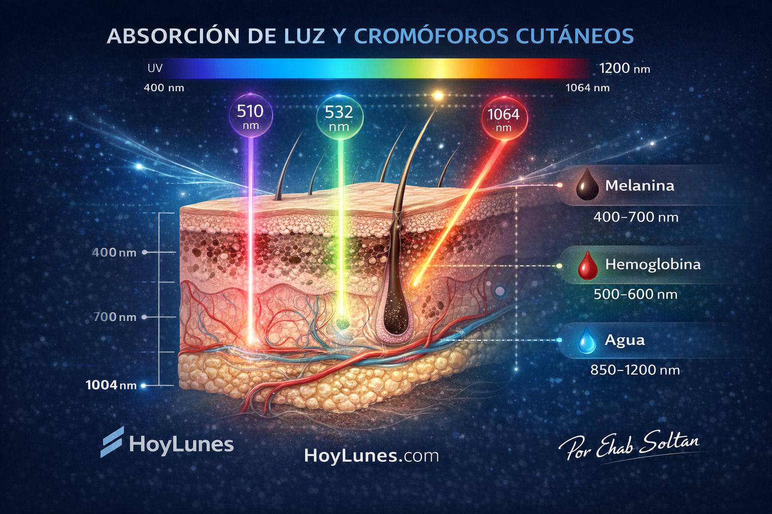
Absorption Engineering and its Targets
Every component of our skin, called a “chromophore”, has a “favorite color” of light that it absorbs avidly. The mastery of the photonic dermatologist lies in choosing the right tool for each objective:
Melanin: The target in pigmented lesions and tattoo removal.
Hemoglobin: The key to treating rosacea, angiomas, and vascular lesions.
Water: The vehicle for deep dermal remodeling and resurfacing.
Biophotonics: Beyond Heat
The current frontier does not seek to burn, but to “speak” to the cells. This is where **photobiomodulation** emerges. By using low-intensity light, it is possible to stimulate the mitochondria (specifically the cytochrome c oxidase enzyme) to accelerate healing, reduce chronic inflammation, and improve ATP synthesis. It is, quite literally, charging our cells’ batteries with photons.
Recent studies show that photobiomodulation activates intracellular pathways such as NF-κB, modulates reactive oxygen species (ROS) at non-cytotoxic levels, and alters intracellular calcium gradients. These responses are photochemical rather than thermal. The result is a transcriptional modulation that impacts inflammation, angiogenesis, and tissue regeneration. Light, in this context, acts as a biological signal and not a destructive agent.
The Therapeutic Arsenal: From Heat to Acoustic Impact
The evolution of devices has allowed for milestones that were previously unthinkable:
Picosecond Lasers: If the traditional laser was a “hot hammer,” the picosecond laser is an ultrasonic shockwave. By firing in $10^{-12}$ seconds, it fragments pigment through a photoacoustic effect, removing tattoos and spots with near-zero thermal damage. This paradigm shift—from photothermal to photomechanical—significantly reduces post-inflammatory inflammation and the risk of hyperpigmentation in high phototypes.
Photodynamic Therapy (PDT): A strategic alliance between chemistry and light. A sensitizing agent is applied to precancerous cells; when illuminated, they die selectively, sparing healthy tissue. This is the spearhead against actinic keratosis and certain carcinomas.
Next-Generation Intense Pulsed Light (IPL): Thanks to smart filters, modern IPL allows for the treatment of multiple conditions (spots and veins) in a single session with optimized safety profiles.

The End of the Scalpel: The Era of the “Optical Biopsy
Perhaps the most respected advancement by the academic community is the ability to “see” beneath the skin without the need for cutting.
In Vivo Confocal Microscopy: This device allows specialists to navigate the layers of the epidermis in real-time, visualizing living cells with a resolution that rivals classical histology. It is a virtual biopsy: instantaneous and scarless.
Optical Coherence Tomography (OCT): Using interferometry, OCT generates three-dimensional maps of the skin’s architecture, allowing for the surgical-precision monitoring of tumor margins before operating.
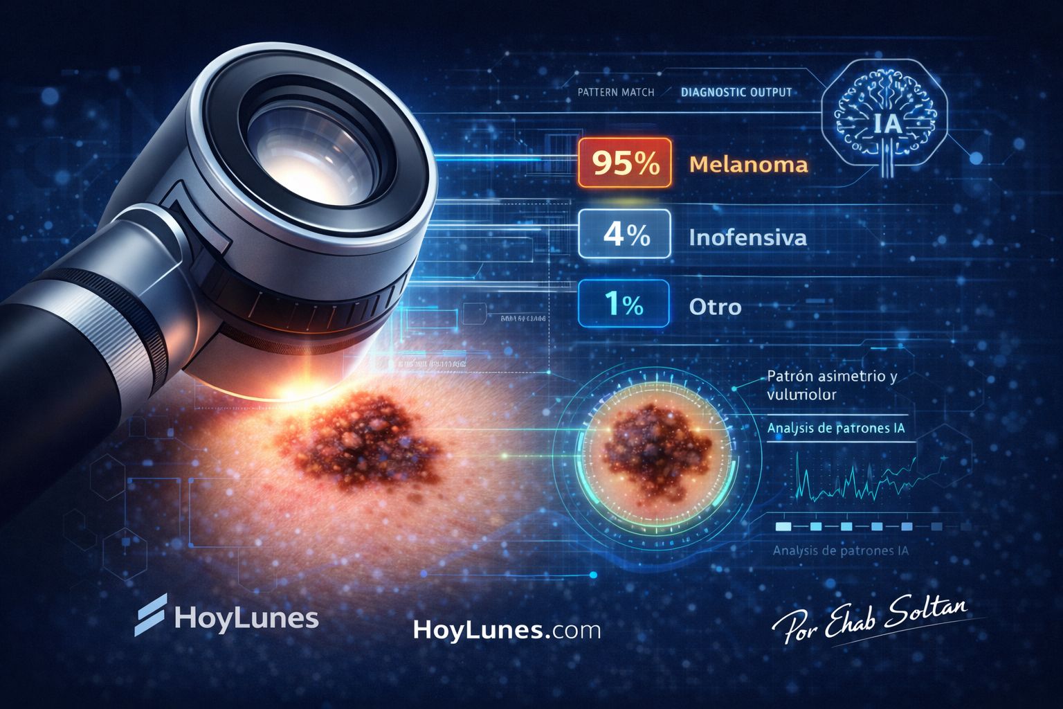
AI + Photonics: Diagnosis no longer depends solely on the human eye. Deep learning algorithms analyze the light signatures of lesions, helping to detect melanomas at such early stages that survival rates soar above 95%.
The true revolution lies not only in diagnostic capability but in parametric personalization. Emerging systems combine spectral analysis, Fitzpatrick phototype, vascular density, and lesion depth to adjust wavelength, fluence, and pulse duration in real-time. Dermatology thus enters the era of adaptive treatment assisted by algorithms, where human variability ceases to be an obstacle and becomes an optimizable data point.
Ethics, Safety, and Quality of Life
The democratization of technology carries risks. The proliferation of “light-based” home devices without medical supervision is a growing concern. Misapplied photonics can mask skin cancer behind an aesthetic appearance or cause irreversible damage.
Photonic medicine is not a “salon” treatment; it demands rigorous training in medical optics and physiology. Technology does not replace clinical judgment; it empowers it.
Clinical Impact on Chronic and Oncological Diseases
In rosacea, photonics allows for the selective coagulation of microvasculature without compromising the skin barrier. In severe inflammatory acne, the targeted destruction of *Cutibacterium acnes* using endogenous porphyrins reduces bacterial load without systemic antibiotics. In psoriasis, localized immune modulation avoids systemic immunosuppression.
In cutaneous oncology, confocal microscopy and OCT are reducing unnecessary biopsies and improving pre-surgical tumor delimitation. The consequence is not merely aesthetic: it is a reduction in morbidity, shorter surgical times, and the optimization of healthcare resources.
Illuminating Before Cutting
Dermatology has transitioned from indiscriminate ablation to intelligent intervention. Light is no longer just energy; it is pure information. If the 20th century was the century of steel and the scalpel, the 21st century indisputably belongs to the photon.
At HoyLunes, we understand that Photonic Medicine is the perfect convergence of quantum physics and clinical empathy. The future of health will not come from deeper cuts, but from more precise light. Because in the medicine of the future, the first step will not be to cut; it will be to illuminate.
In the next decade, we will see hybrid devices integrating fractional lasers, confocal diagnosis, and AI-automated adjustment into a single platform. The miniaturization of solid-state laser sources and integration with portable spectroscopic sensors will broaden hospital and outpatient access. Photonic dermatology will not only be more precise; it will be more integrated, predictive, and personalized.
Scientific Sources and Technical References
Anderson RR, Parrish JA. Selective photothermolysis: precise microsurgery by selective absorption of pulsed radiation. Science (1983).
[DOI: 10.1126/science.6836297]
Jacques SL. Optical properties of biological tissues: a review. Phys Med Biol (2013). (https://iopscience.iop.org/article/10.1088/0031-9155/58/11/R37)
Hamblin MR. Mechanisms and applications of the anti-inflammatory effects of photobiomodulation. AIMS Biophysics (2017). [PMC5523874]
Esteva A, et al. Dermatologist-level classification of skin cancer with deep neural networks. Nature (2017). [Nature 21056]
Avci P, et al. Low-level laser (light) therapy in skin: stimulating, healing, restoring. Semin Cutan Med Surg (2013). [PMC4126803]
González S, et al. In vivo reflectance-mode confocal microscopy in dermatology. J Am Acad Dermatol (1999). [https://pubmed.ncbi.nlm.nih.gov/10459113/]
`#PhotonicMedicine` `#ModernDermatology` `#HoyLunes` `#Biophotonics` `#DigitalHealth2026` `#BiomedicalEngineering` `#SkinCancer` `#MedTech` `#EhabSoltan`